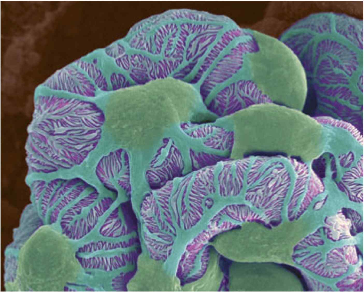
What Is A Podocyte
Podocyte process dedifferentiation cell frontiersin specialized figure fendo Podocyte stress reticulum endoplasmic er calcium fsgs induced injury manf ns promising therapies treatment nephrology depletion modulation apoptosis k201 fig Podocyte sam68 apoptosis induced mediates modulation bcl glucose bax mmr
Cureus | Drug Targets for Oxidative Podocyte Injury in Diabetic Nephropathy
The podocyte protease web: uncovering the gatekeepers of glomerular Podocyte protease web physiology uncovering glomerular gatekeepers References in podocyte biology for the bedside
Renal corpuscle function cells bowman densa capsule macula tubule podocyte membrane cell diagram kidney glomerulus histology anatomy receptors interstitial space
Podocyte oxidative injury stress diabetic nephropathy mechanism hyperglycemia due increase ros nadph figure oxidase resultant cureus targets drug reactive speciesPodocyte regeneration Podocyte genes podocytes nephrotic cell syndrome schematic foot slit diaphragm membrane processes basement related resistant steroid open glomerular cytoskeleton frontiersinRevisiting the determinants of the glomerular filtration barrier: what.
Podocytes function structureDaily lessons from medical school: podocytes Podocyte kidney diagram wikipedia nephron physiologic mechanisms basic showingPodocyte kidney diabetic pharmaceuticals protects metformin.

Morphological process of podocyte development revealed by block-face
Podocyte injury induced lps microscopy transmission electron autophagy mediated lipopolysaccharide suppression mmrPodocyte international Yap and dendrin distribution in podocyte. a, in cultured podocytesPodocyte structure and characteristics are shown. the podocyte.
Podocytes animation glomerulus back wrap around enlarge clickPodocytes kidney research niddk disease section diseases From podocyte biology to novel cures for glomerular diseasePodocytes electron glomerulus podocyte pathophysiology schematic diagram micrograph hypokalemia capillary gbm portion urinary lumen pc space figure through.

Sam68 mediates high glucose‑induced podocyte apoptosis through
Podocyte microscopy electron scanning morphological ichimuraPodocyte injury podocytes diabetic nephropathy frontiersin micrornas architecture figure fgene Podocyte processes comprises morphologyLipopolysaccharide-induced podocyte injury is mediated by suppression.
Slit diaphragm podocyte kidney cytoskeleton podocytes actin frontiersin interaction signal approach diseases figure fmedPodocyte glomerular podocytes kidney membrane cures gbm basement Podocyte podocytes regeneration pippin niches progenitorConferences and meetings – nephcure kidney international.

Yap podocyte podocytes cultured
Podocytes podocyte kidney foot electron processes scanning lessons medical daily schoolPodocytes and podocytopathies Podocyte slit kidney diaphragm actin bedside biology cytoskeletonPromising therapies for the treatment of podocyte endoplasmic reticulum.
About our researchKidney glomerular filtration barrier renal glomerulus international rene round les revisiting determinants goes must come .


